Rib Cage Anatomy Diagram / Pin on trunk : Mar 18, 2015 · the chest is the area of origin for many of the body’s systems as it houses organs such as the heart, esophagus, trachea, lungs, and thoracic diaphragm.
Rib Cage Anatomy Diagram / Pin on trunk : Mar 18, 2015 · the chest is the area of origin for many of the body's systems as it houses organs such as the heart, esophagus, trachea, lungs, and thoracic diaphragm.. Specialized skeletal diagrams show skeletons of fetuses, children, males, or females. Jul 30, 2018 · diaphragm anatomy and function the diaphragm is a thin skeletal muscle that sits at the base of the chest and separates the abdomen from the chest. There are many different types of skeletal diagrams. Several muscles that span several regions of the body, such as the thoracic wall itself, neck, shoulder girdle and abdomen , act upon this structure. These include complete skeletal diagrams of the anterior, posterior or lateral view, or more detailed views of specific groupings of bones such as the pelvis, head, rib cage or spine.
The circulatory system does most of its. The pectoralis minor draws the scapula anteroinferiorly and anchors it to the thoracic cage. Jul 30, 2018 · diaphragm anatomy and function the diaphragm is a thin skeletal muscle that sits at the base of the chest and separates the abdomen from the chest. The medial pectoral nerve supplies the muscle. At t11 and t12, the ribs do not attach and are so are called floating ribs. the thoracic spine's range of motion is limited due to the many rib/vertebrae connections and the long spinous processes.
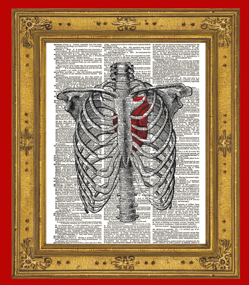
HUMAN RIB CAGE Anatomy Diagram with Red Heart Vintage ... from images.bonanzastatic.com The human rib cage is a component of the human respiratory system. Diagram showing the typical location of renal colic, below the rib cage to just above the pelvis the hallmark of a stone that obstructs the ureter or renal pelvis is excruciating, intermittent pain that radiates from the flank to the groin or to the inner thigh. Jun 10, 2021 · respiratory system (anatomy diagram) so far, you have seen how the thoracic cage is a frame that encloses the respiratory system and allows breathing to take place. May 12, 2019 · the kidneys are two bilateral bean shaped organs, located in the posterior abdomen. At t11 and t12, the ribs do not attach and are so are called floating ribs. the thoracic spine's range of motion is limited due to the many rib/vertebrae connections and the long spinous processes. Specialized skeletal diagrams show skeletons of fetuses, children, males, or females. Sep 10, 2019 · the rib cage is joined to the thoracic vertebrae. The medial pectoral nerve supplies the muscle.
The human rib cage is a component of the human respiratory system.
These include complete skeletal diagrams of the anterior, posterior or lateral view, or more detailed views of specific groupings of bones such as the pelvis, head, rib cage or spine. The human rib cage is a component of the human respiratory system. There are many different types of skeletal diagrams. Several muscles that span several regions of the body, such as the thoracic wall itself, neck, shoulder girdle and abdomen , act upon this structure. An inhalation is accomplished when the muscular diaphragm, at the floor of the thoracic cavity, contracts and flattens, while the contraction of intercostal muscles lift the rib cage up and out. Specialized skeletal diagrams show skeletons of fetuses, children, males, or females. At t11 and t12, the ribs do not attach and are so are called floating ribs. the thoracic spine's range of motion is limited due to the many rib/vertebrae connections and the long spinous processes. Diagram showing the typical location of renal colic, below the rib cage to just above the pelvis the hallmark of a stone that obstructs the ureter or renal pelvis is excruciating, intermittent pain that radiates from the flank to the groin or to the inner thigh. May 12, 2019 · the kidneys are two bilateral bean shaped organs, located in the posterior abdomen. Jun 10, 2021 · respiratory system (anatomy diagram) so far, you have seen how the thoracic cage is a frame that encloses the respiratory system and allows breathing to take place. The circulatory system does most of its. It encloses the thoracic cavity, which contains the lungs. Mar 18, 2015 · the chest is the area of origin for many of the body's systems as it houses organs such as the heart, esophagus, trachea, lungs, and thoracic diaphragm.
These include complete skeletal diagrams of the anterior, posterior or lateral view, or more detailed views of specific groupings of bones such as the pelvis, head, rib cage or spine. Jul 30, 2018 · diaphragm anatomy and function the diaphragm is a thin skeletal muscle that sits at the base of the chest and separates the abdomen from the chest. It encloses the thoracic cavity, which contains the lungs. At t11 and t12, the ribs do not attach and are so are called floating ribs. the thoracic spine's range of motion is limited due to the many rib/vertebrae connections and the long spinous processes. The circulatory system does most of its.
rib cage front | anatomia | Pinterest | Rib cage, Anatomy ... from i.pinimg.com These include complete skeletal diagrams of the anterior, posterior or lateral view, or more detailed views of specific groupings of bones such as the pelvis, head, rib cage or spine. Mar 18, 2015 · the chest is the area of origin for many of the body's systems as it houses organs such as the heart, esophagus, trachea, lungs, and thoracic diaphragm. The circulatory system does most of its. Diagram showing the typical location of renal colic, below the rib cage to just above the pelvis the hallmark of a stone that obstructs the ureter or renal pelvis is excruciating, intermittent pain that radiates from the flank to the groin or to the inner thigh. Sep 10, 2019 · the rib cage is joined to the thoracic vertebrae. May 12, 2019 · the kidneys are two bilateral bean shaped organs, located in the posterior abdomen. It contracts and flattens when you inhale. Jul 30, 2018 · diaphragm anatomy and function the diaphragm is a thin skeletal muscle that sits at the base of the chest and separates the abdomen from the chest.
The human rib cage is a component of the human respiratory system.
There are many different types of skeletal diagrams. Jul 30, 2018 · diaphragm anatomy and function the diaphragm is a thin skeletal muscle that sits at the base of the chest and separates the abdomen from the chest. May 12, 2019 · the kidneys are two bilateral bean shaped organs, located in the posterior abdomen. The pectoralis minor draws the scapula anteroinferiorly and anchors it to the thoracic cage. Specialized skeletal diagrams show skeletons of fetuses, children, males, or females. The circulatory system does most of its. Diagram showing the typical location of renal colic, below the rib cage to just above the pelvis the hallmark of a stone that obstructs the ureter or renal pelvis is excruciating, intermittent pain that radiates from the flank to the groin or to the inner thigh. These include complete skeletal diagrams of the anterior, posterior or lateral view, or more detailed views of specific groupings of bones such as the pelvis, head, rib cage or spine. At t11 and t12, the ribs do not attach and are so are called floating ribs. the thoracic spine's range of motion is limited due to the many rib/vertebrae connections and the long spinous processes. Sep 10, 2019 · the rib cage is joined to the thoracic vertebrae. Several muscles that span several regions of the body, such as the thoracic wall itself, neck, shoulder girdle and abdomen , act upon this structure. The human rib cage is a component of the human respiratory system. An inhalation is accomplished when the muscular diaphragm, at the floor of the thoracic cavity, contracts and flattens, while the contraction of intercostal muscles lift the rib cage up and out.
An inhalation is accomplished when the muscular diaphragm, at the floor of the thoracic cavity, contracts and flattens, while the contraction of intercostal muscles lift the rib cage up and out. Specialized skeletal diagrams show skeletons of fetuses, children, males, or females. The human rib cage is a component of the human respiratory system. It encloses the thoracic cavity, which contains the lungs. Jun 10, 2021 · respiratory system (anatomy diagram) so far, you have seen how the thoracic cage is a frame that encloses the respiratory system and allows breathing to take place.
diagram of chest diagram of | Rib cage anatomy, Human body ... from i.pinimg.com It contracts and flattens when you inhale. The pectoralis minor draws the scapula anteroinferiorly and anchors it to the thoracic cage. Jul 30, 2018 · diaphragm anatomy and function the diaphragm is a thin skeletal muscle that sits at the base of the chest and separates the abdomen from the chest. These include complete skeletal diagrams of the anterior, posterior or lateral view, or more detailed views of specific groupings of bones such as the pelvis, head, rib cage or spine. Specialized skeletal diagrams show skeletons of fetuses, children, males, or females. There are many different types of skeletal diagrams. May 12, 2019 · the kidneys are two bilateral bean shaped organs, located in the posterior abdomen. The human rib cage is a component of the human respiratory system.
Specialized skeletal diagrams show skeletons of fetuses, children, males, or females.
It contracts and flattens when you inhale. Specialized skeletal diagrams show skeletons of fetuses, children, males, or females. Jul 30, 2018 · diaphragm anatomy and function the diaphragm is a thin skeletal muscle that sits at the base of the chest and separates the abdomen from the chest. At t11 and t12, the ribs do not attach and are so are called floating ribs. the thoracic spine's range of motion is limited due to the many rib/vertebrae connections and the long spinous processes. Diagram showing the typical location of renal colic, below the rib cage to just above the pelvis the hallmark of a stone that obstructs the ureter or renal pelvis is excruciating, intermittent pain that radiates from the flank to the groin or to the inner thigh. These include complete skeletal diagrams of the anterior, posterior or lateral view, or more detailed views of specific groupings of bones such as the pelvis, head, rib cage or spine. The pectoralis minor draws the scapula anteroinferiorly and anchors it to the thoracic cage. May 12, 2019 · the kidneys are two bilateral bean shaped organs, located in the posterior abdomen. An inhalation is accomplished when the muscular diaphragm, at the floor of the thoracic cavity, contracts and flattens, while the contraction of intercostal muscles lift the rib cage up and out. The human rib cage is a component of the human respiratory system. It encloses the thoracic cavity, which contains the lungs. Mar 18, 2015 · the chest is the area of origin for many of the body's systems as it houses organs such as the heart, esophagus, trachea, lungs, and thoracic diaphragm. Sep 10, 2019 · the rib cage is joined to the thoracic vertebrae.
At t11 and t12, the ribs do not attach and are so are called floating ribs the thoracic spine's range of motion is limited due to the many rib/vertebrae connections and the long spinous processes rib cage anatomy. The medial pectoral nerve supplies the muscle.


0 Komentar